Skip to content
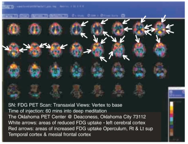
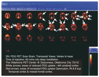
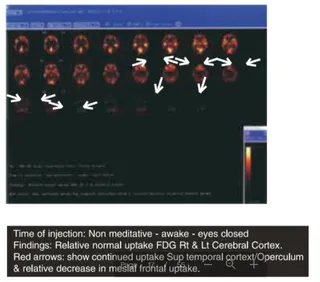
A screenshot of a medical imaging viewer showing a grid of colour-coded transaxial PET brain scans arranged in rows, from vertex to base. White arrows point to areas of reduced FDG uptake in the left cerebral cortex, and red arrows point to areas of increased uptake in the operculum, right and left superior temporal cortex and mesial frontal cortex. A caption on the lower left reads the scan was taken 60 minutes into deep meditation at the Oklahoma PET Center. No people are visible; the scan slices are brain images, not physical people.