Skip to content
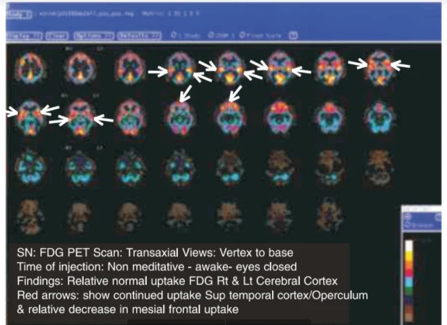
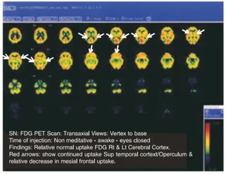
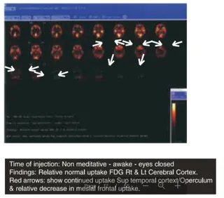
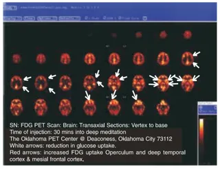
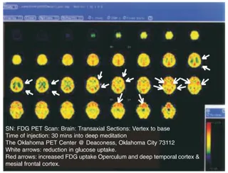
A screenshot of a medical imaging viewer showing a grid of colourful FDG PET brain scan slices in transaxial views, from vertex to base, displayed in orange, yellow, red, blue and teal. Red arrows point to highlighted regions in the upper slices. A caption box overlaid at the bottom describes the scan: non-meditative, awake with eyes closed, relative normal uptake in the right and left cerebral cortex, continued uptake in the superior temporal cortex and operculum, and a relative decrease in mesial frontal uptake. A colour scale bar sits at the right edge. No people are visible.
Photo 2 of 6 from 10 March 2004