Skip to content
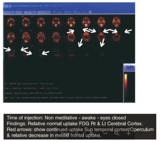
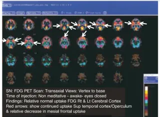
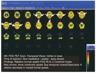
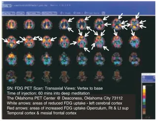
A screenshot of a brain imaging viewer showing a grid of axial PET scan slices on a dark background, displayed in false colour with orange, red and yellow hot spots indicating FDG uptake. White double-headed arrows point to regions of the scan, and a colour scale bar sits at the lower right. Below the image, a black caption bar states the time of injection as non-meditative, awake with eyes closed, and notes relative normal uptake in the right and left cerebral cortex, continued uptake in the superior temporal cortex and operculum, and a relative decrease in mesial frontal uptake. The slide belongs to a page on research conclusions about the brain.