Skip to content
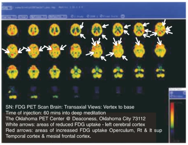
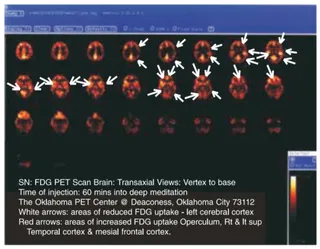
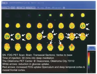
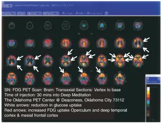
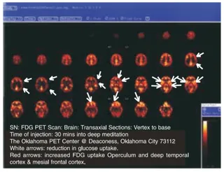
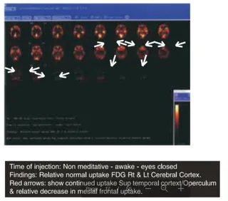
A screenshot-style scientific figure showing a grid of colour-mapped transaxial PET brain scans, arranged in rows from vertex to base on a dark background. The brain slices are rendered in yellow, orange, red, green and blue, with white arrows pointing to areas of reduced uptake and red arrows marking areas of increased uptake. Software toolbar text runs across the top of the image and a colour intensity scale appears at the lower right. A caption block at the bottom describes the FDG PET brain scan, its injection timing and the imaging centre. No people are visible; the image depicts brain scan data only.
Photo 4 of 6 from 10 March 2004