Skip to content
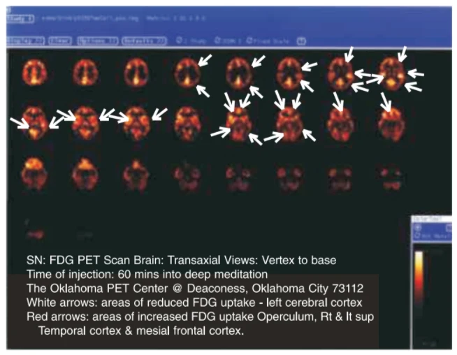
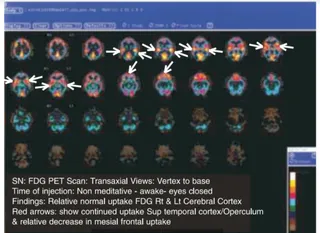
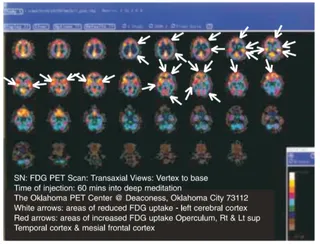
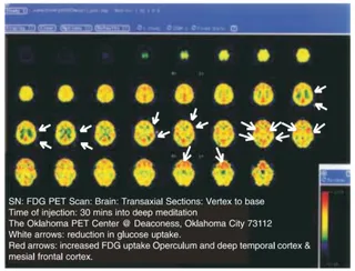
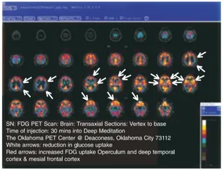
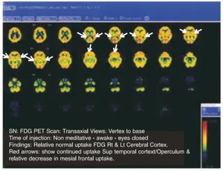
A screenshot of a PET brain scan display titled as an FDG PET scan, showing a grid of transaxial brain slices in orange, yellow and red on a black background, with white and red arrows marking areas of reduced and increased FDG uptake. A dark caption box at the bottom lists the scan details, and a colour scale bar sits at the lower right. No people are shown.