Skip to content
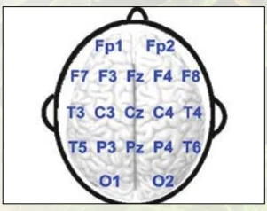
A graphic diagram of a human brain viewed from above, drawn in grey with a white and grey cortical surface inside a black oval outline. Blue electrode position labels are arranged across the scalp in rows: Fp1 and Fp2 at the front, F7, F3, Fz, F4, F8 beside them, T3, C3, Cz, C4, T4 at the sides, T5, P3, Pz, P4, T6 further back, and O1 and O2 at the rear. This is an EEG electrode placement chart, with no people shown.
Photo 282 of 290 from 12 April 2013