Skip to content
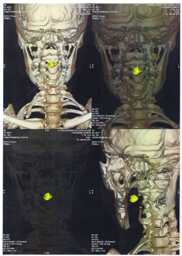
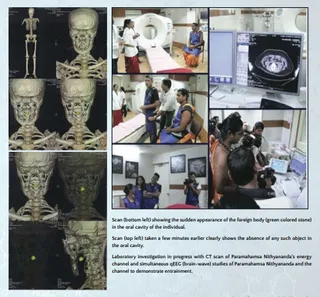
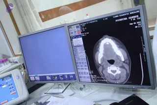
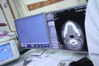
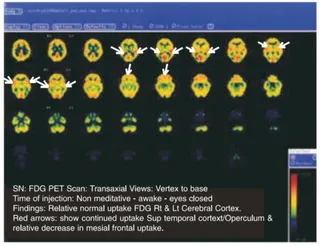
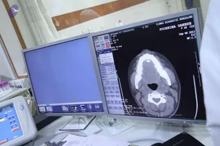
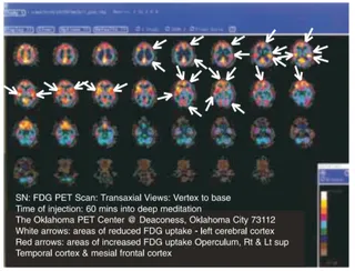
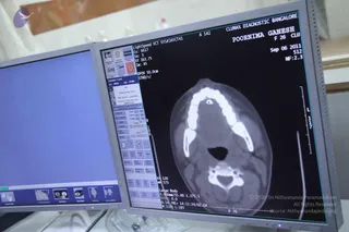
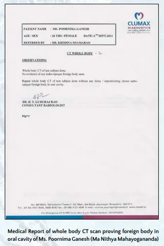
A four-panel scan of 3D volume-rendered CT images of a human neck and skull, shown in grey and bone-toned shades against black backgrounds. Each panel shows the cervical spine, jaw and clavicles from front and side views, with a bright yellow highlighted mass or marker at the upper cervical spine. Printed header and footer details identify the diagnostic centre, a patient name and dates, and the imaging parameters. No people are physically present; the subject is an anatomical scan.